From November 2019 to January 2020, two hundred Clinical specimens (Wound, Burns swabs and Urine samples) were collected from patients admitted to Baquba Teaching Hospital and Al-Batoul Hospital. Samples were cultured on MacConkey and Blood agar media. The bacterial isolates were then initially diagnosed using selective and differential media. Then biochemical tests were performed to confirm the diagnose of bacterial isolates. Based on the biochemical identification the bacterial spp. were as followings: Klebsiella pneumoniae, Escherichia coli, Proteus mirabilis, Pseudomonas aeruginosa, Staphylococcus epidermidis and Staphylococcus aureus.
Antimicrobial susceptibility test, for all isolated bacterial species were evaluated toward 29 antimicrobial agents using disk diffusion method. The results showed that many bacterial isolates were multiple drugs resist (MDR).
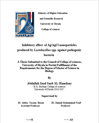
Biosynthesis method of nanoparticles acquires very important area due to their economic and ecofriendly benefits. In the present study, used cell free supernatant of lactobacillus spp. for synthesis of silver nanoparticles. Lactobacillus spp. bacteria were isolated from local fermented product and identified as Lactobacillus spp. by using selective enrichment culture medium de Man, Rogosa and Sharpe agar (MRS agar). Morphology and microscopical diagnose and biochemical tests were used to confirm the identification of Lactobacillus . The cell free supernatants was used for biosynthesis of Silver nanoparticales.
The silver nanoparticles were Characterized using X-ray diffraction (XRD) analysis to confirm that the nanoparticles were silver and silver chloride (AgCl) cubic types. The (crystalline) domains equation, showed three strongest peaks, the calculated particle size were 17.1and 18.6 nm for Ag, and 24.3 nm for AgCl. Atomic Force Microscopy (AFM) used to characterize the size, topography and granularity volume distribution of biosynthesized nanoparticles Ag/AgCl, average size was 50 nm. UV-visible spectroscopy revealed the formation of AgNPs. Fourier transform infrared (FTIR) spectroscopy showed different functional groups of biomolecules which were responsible for reduction and capping process. Electron microscopic (SEM) was used to characterize the shape and size of the synthesized nanoparticles.
The antibacterial activity of silver and sliver chloride nanoparticles against the selected multiple drugs resist (MDR) bacteria were determined by agar well diffusion method. It was observed that the growth of these bacteria was inhibited at 12,500 µg /ml of Ag&AgCl NPs. The nanoparticles concentrations were (12,500 ; 25,000 ; 50,000 ; 100,000) µg/ml had maximum inhibitory effect against S. aureus (24, 28, 30, 32 mm) respectively and minimum inhibitory effect against E. coli (16 , 21, 23, 27 mm) respectively while the others bacteria (P. mirabilis, S. epidermidis, K. pneumoniae and P. aeruginosa) inhibitory effect between the bacterial isolates and the concentration of the silver nanoparticles.
Finally, determination of MIC of Ag/AgCl nanoparticles was done by microdilution method. The result showed the MIC for Ag/AgCl NPs for P. mirabilis, S. aureus, S. epidermidis and P. aeruginosa was 2500 µg/ml while the MIC for E. coli, K. pneumoniae was 5000 µg/ml.

